Caribbean Institute of Endodontics

Trusted by Patients Across Jamaica

Articles

Successful endodontic treatment involves accurate diagnosis, good understanding of the biological principles and excellent execution of the treatment. To be able to execute an excellent treatment, it’s imperative that the clinician has comprehensive knowledge of the root canal anatomy and the know-how to locate and treat this anatomy.

Many outcome studies conducted over the past few decades showed incomplete debridement and disinfection of root canal space as the most important factor in endodontic treatment failure (1,2,3). Missed canal in the initial treatment as a significant cause of root canal failure was shown by Hoen et al (4). They also showed a significant relation between an asymmetrical obturation in a root space and the incidence of a missed canal in an initial treatment.

Methodology

Root canal anatomy is studied by both in vitro and in vivo methods. In vivo methods include clinical treatment of a tooth followed by radiographic evaluation of the root canal anatomy. In vitro methods include:

  1. Direct observation
  2. Microscopic observation
  3. Macroscopic sectioning
  4. Microscopic sectioning
  5. Dyes
  6. Filling and decalcification
  7. Filling and clearing
  8. Radiography
  9. Contrasting media (Hypaque)
  10. Cone beam Tomography

Classification

Weine classified root canal anatomy into 3 types:

  • Type I: One canal with one orifice and one apical foramen (1-1)
  • Type II: Two canals that merge into one and exit as one canal (2-1)
  • Type III: One canal that divides into two and exit as two canals. (2-2)

Vertucci’s classification was more elaborate and it covered 8 types:

  • Type I: 1-1
  • Type II: 2-1
  • Type III: 1-2-1
  • Type IV: 2-2
  • Type V: 1-2
  • Type VI: 2-1-2
  • Type VII: 1-2-1-2
  • Type VIII: 3-3

Studies with Percentages

The methodology employed and the criteria used to describe root canal anatomy will decide the percentages of canals found in any tooth. For example, Neaverth et al (5) have used “two separate canals could be visualized on radiographic examination (two files or two GP points to no less than a mm short of the length)” as the criteria for two canals in a mesiobuccal root of a maxillary first molar. On the other hand, Sempira et al (6) have used the presence of “two canals to at least 4 mm from the apex” in the mesiobuccal root of maxillary first molar to determine the percentage of Mb2 canal.

These differences in criteria dictate the great variation seen in the percentages of root canals seen in different studies. However, if one reviews the published evidence, certain features of aberrant anatomy stand out in a tooth/root.

  • Ethnicity has a significant influence on aberrant anatomy (26). Radix Entomolaris, an extra distal root in a mandibular molar, is often seen in Oriental and Eskimo populations (23). Similarly, 2 and 3 canal premolars are seen frequently in black populations (12,15,27). ‘C’ shaped anatomy is seen more commonly in Chinese, Korean and Indian populations (22,25).
  • Bilateral symmetry is a feature of aberrant anatomy. Rarer the aberration, the more common is the bilateral symmetry (28).

Clinical Management of Aberrant Anatomy

Radiography: Angled views of teeth reveal aberrant anatomy. Angled views allow us to visualize the root anatomy in 3 dimensions so that better assessment of the root canal anatomy is made. It is imperative that at least 2 angled views shall be taken before attempting endodontic treatment.

Author Profile

Dr. Sashi Nallapati obtained his dental degree from the Govt. Dental College and Hospital, Hyderabad, India. He completed his post graduate training in the specialty of Endodontics from Nova Southeastern University (NSU), Davie, Florida, USA. He maintains a practice limited to Endodontics in Kingston, Jamaica. He serves on the faculty of NSU in the dept. of Post Graduate Endodontics. Dr. Nallapati authored several clinical articles and lectures across the globe. His hobbies include digital photography, swimming and reading. He can be reached atwww.endojamaica.com.

Abstract

Mandibular premolars have earned the reputation for having aberrant anatomy. The occurrence of three canals with three separate (type V, Vertucci) foramina in mandibular premolars is very rare. If one is to treat mandibular premolar teeth with three canals predictably, it is necessary to be aware of their clinical and radiographic anatomy. These teeth may also require special shaping and obturating techniques. This article reports and discusses the treatment recommendations for an unusual occurrence of three canals with three separate foramina in both the first and second mandibular premolars in the same patient.

Consistent, high levels of success in endodontic treatment require an understanding of root canal anatomy and morphology. To achieve endodontic success, the entire root canal system must be debrided, disinfected and obturated. The clinician must have a thorough understanding of normal anatomy, and of common variations from the norm. The clinician must also be prepared to identify those teeth that tend to vary greatly from the norm, e.g. mandibular premolars.

Quite a few studies were published in endodontic literature that studied the common reasons for endodontic failures (1–3). Hoen and Pink found a 42% incidence of missed roots or canals in the teeth that needed retreatment in their investigations (4). They concluded that the clinical application of a thorough knowledge of canal anatomy and meticulous attention to treatment detail are essential to minimizing failure and the need for subsequent endodontic retreatment.

In a series of studies on extracted teeth Vertucci in the 1970s and 1980s determined canal numbers and configurations by percentages for each of the teeth (5). Vertucci’s data may not be exactly representative of different locations and ethnic groups, but it is a good starting point for understanding root canal anatomy.

Mandibular premolars have gained a reputation for having aberrant anatomy. Different studies have looked at the root canal morphology of mandibular premolars over the years and reported a fairly high percentage of these teeth to have more than one canal (6–9).

There seems to be a racial predisposition for the presence of two or more canals in maxillary and mandibular premolars (10, 11), as well as their bilateral occurrence (12). The occurrence of three canals with three separate (type V, Vertucci) foramina in mandibular premolars is very rare. Vertucci and Zillich et al. reported the occurrence of three canals in mandibular first premolars at 0.5% and 0.4% respectively. Their studies in second premolars showed these percentages at 0.0% and 0.4%, respectively (13, 14).

If one is to treat mandibular premolar teeth with three canals, predictably, it is necessary to be aware of their clinical and radiographic anatomy. These teeth may also require special shaping and obturating techniques.

The purpose of this article is to report, as well as discuss, treatment recommendations for an unusual occurrence of three canals with three separate foramina in both the first and second mandibular premolars in the same patient.

Case Report

A 49 yr-old White Jamaican male with a noncontributory medical history was referred to the author’s private practice for endodontic treatment on the right mandibular first and second premolars by his general dentist.

The chief complaint of the patient was “pain in the lower right back teeth.” Clinical examination revealed restorations in the right mandibular first and second premolar teeth (28, 29) with distal caries in 29. Teeth 30 and 31 were missing and 32 drifted mesially. Teeth 28 and 29 tested percussion sensitive. Vitality tests on both teeth showed no response to cold and EPT. Radiographic examination revealed periradicular radiolucencies in relation to 28 and 29. More than one root canal was suspected in both the premolars (Fig. 1A).

A pulpal diagnosis of necrotic pulp and a periradicular diagnosis of acute exacerbation of a chronic periradicular periodontitis were made. Nonsurgical endodontic treatment was planned in both 28 and 29 over two visits with the use of calcium

A maxillary right lateral incisor with a type 3 dens invaginatus and a large periapical lesion with vital pulp in a separate root canal was treated both nonsurgically and surgically. Care was taken not to expose or devitalize the vital pulp in the main root canal system during the treatment. The signs and symptoms ceased after the treatment, and 4-month recall showed complete bone healing with pulp vitality maintained.

Dens invaginatus is a rare malformation of teeth with a broad spectrum of morphological variations. The affected teeth present with an infolding of enamel and dentin, which may extend into the pulp cavity, into the root, and sometimes to the root apex (1). Several theories have been proposed for this phenomenon, but the etiology of dens invaginatus remains unclear. Kronfeld (2) proposed that dens invaginatus is caused by a focal failure of growth of the internal enamel epithelium leading to proliferation of the surrounding normal epithelium with eventual engulfment of the static area. Oehlers (3) proposed that distortion of the enamel organ occurs during tooth development and results in protrusion of a part of the enamel organ. Other theories include infection (4), trauma (5), and genetics (6) as possible contributing factors.

Oehlers (3) classified these malformations into three types. Of particular interest in his classification is type III, in which an enamel and/or cementum-lined dens forms tracks through the root and perforates in the apical area to form a second foramen but has no direct communication with the pulp. The purpose of this article is to discuss the clinical management of this particular variant of the dens invaginatus form.

CASE REPORTS

A healthy 24-year-old man was referred to the author’s private practice with the chief complaint of a gum boil above the upper right front tooth. The patient had no significant medical history.

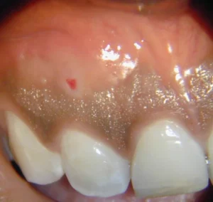
Clinical examination revealed an intraoral sinus tract in the labial gingiva adjacent to the maxillary right lateral incisor (Fig. 1). Clinical tests revealed the maxillary right lateral incisor to be within normal limits to percussion and slightly tender to palpation, with a normal response to cold. The vitality of the pulp was later confirmed with a test cavity. Radiographic examination revealed a $1 \times 2\text{ cm}$ radiolucency adjacent to the mesial aspect of the apex of the maxillary right lateral incisor (Fig. 2). The sinus tract was traced with a gutta-percha point to the lesion.

Radiographs revealed an enamel-lined tract, mesial to and separate from the main root canal system, that tracked to the apical radiolucency and appeared to be wide-open at the portal of exit. There was a separate root canal system distal to the dens that appeared to be closed apically. A diagnosis was made of normal pulp with chronic apical periodontitis associated with the type 3 dens invaginatus.

A treatment plan was formulated that included nonsurgical endodontic treatment of the dens tract over several appointments, including placement of a calcium hydroxide dressing. Surgical intervention was considered a strong possibility. Every effort would be made to leave the main root canal system undisturbed.

After a test cavity, which confirmed the vitality of pulp, the tooth was anesthetized, and nonsurgical endodontic treatment was initiated. All procedures were performed with the aid of a surgical operating microscope. Of particular interest was the atypical presentation of the lingual surface of the tooth. A rubber dam was placed, and access was made in the mesiolingual aspect of the tooth (Fig. 3). Upon access into the dens, there was a serosanguinous discharge, which was allowed to drain (Fig. 4). The canal was shaped initially with long tapered diamond bur to improve access to the apical defect. Working length of the dens tract was determined with the help of an apex locator (Root Zx, J Morita). The dens was debrided as well as possible with Gates Glidden drills and hand files. Because the canal was lined with enamel, methods for cleaning and shaping were not very effective. Irrigation was performed with 5.25% sodium hypochlorite, 17% EDTA, and 100% alcohol. Calcium hydroxide (Ultracal, Ultradent, UT) was placed in the dens tract between appointments (Fig. 5). The access cavity was sealed with Cavit and IRM between visits to prevent contamination of the dens canal system.