Caribbean Institute of Endodontics

Trusted by Patients Across Jamaica

Case studies

First page of the Case Report 02/09/2010 newsletterPatient is a 36 YHM.
Reported to his GP with a discolored tooth 26.(LR Lateral Incisor)
Surprisingly he never had any pain or swelling in the area.
The GP diagnosed the tooth as non vital and opened the tooth. There was significant drainage from the tooth. Patient was referred to me for further management.

Here is my management for this type of lesions.

  • 7/2009: Started treatment, obtained a lot of serous drainage, placed CaOH.
  • 9/2009: More serous drainage was obtained, and more CaoH was placed in the canal.
  • 12/2009: Definitely the lesion is shrinking.
  • 1/2010: Canal was found dry. I obturated the tooth. Bleached it internally with Sodium Perborate.
  • 7/2010: AT 6 months recall, 90% osseous healing seen.

Unfortunately RD never saw my recommendations and crowned the tooth. It wasn’t necessary to crown the tooth.

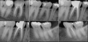
The radiographic series in the case report shows: preoperative images with a larger periapical rarefaction; interim images with intracanal medicament CaOH in the canal, the lesion shrinking in size two months after the initial treatment; further shrinking of the lesion at 5 months; the immediate postoperative view; and the 6 month recall showing excellent healing.

The important take home message for this case is, DO NOT get alarmed by the size of the lesion. Making a correct diagnosis is important before initiating treatment. It was established that it was a Lesion of Endodontic Origin (LEO) from the facts, history of trauma, discoloration of the tooth, RD reporting drainage from tooth. My first reaction was to try the most conservative approach, even though the size of the lesion and the root resorption on 24 made me suspicious to the nature of the etiology. Open the tooth, drain and apply CaOH and wait. If lesion shrinks and canal canal be dried, obturate, and follow up. If lesion does not shrink or canal cannot be dried after multiple CaOH applications, do a surgery and send the specimen to the lab for biopsy to rule out non-odontogenic etiology, or refer to an able Oral Surgeon. I prepared the patient for both RCT and surgery. At the end, I didn’t have to do surgery.

Please remember, time is of essence when you are following up the case. Neoplasias of the jaws can be very aggressive lesions and need to be dealt with immediately. So, an assessment needs to be made if the tooth is responding to non-surgical endodontic treatment or not expediently. Usually, 3 months will be a good period to wait to make that assessment. Referral to an endodontist or an oral surgeon shall be an option for more effective management of the patient.

Please email me at sashi@cwjamaica.com with any comments or questions.

Sashi Nallapati

Download the full case report with all radiographs (PDF)

First page of the Case Report 01/09/2010 newsletterPatient is a 42 YHF. Extremely nervous and scared.
Prior endodontic treatment and apicoectomy in both 7 and 8.
Large lesions that are palpation sensitive.

  • 1/2008: Retreatment started with with long term CaOH.
  • 5/2008: CaOH replaced
  • 8/2008: MTA to seal 8 and Custom fit GP (impression technique) to seal the apex of 7.
  • 3/2010: 19 month recall shows excellent healing.

Her orthodontic treatment is almost complete. She is now changing crowns for both 7,8.

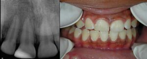
The radiographic series in the case report shows, left to right: Preop — Interim with CaoH — Immediate Postop — 19 Month Recall.

This case highlights that when endodontically treated teeth fail, often, apicoectomy is not the answer. Most RCTs fail because the canal space was not properly cleaned and disinfected in the first treatment. By doing an apicoectomy with or with out a retrofill on these teeth may only give a short term success. In the long term most of these cases may fail.

Quite a few times there is coronal leakage into the canals which contaminates the canal space. By not addressing this contamination with retreatment, and by choosing to do only an apicoectomy, you are risking a late failure. Eventually the contamination gets past the retroseal resulting in a periapical lesion. With newer materials like MTA and time old techniques of Gutta-Percha impression, sealing teeth with blunderbuss /open apex is not a challenging issue any more.

This case shows the clinical handling of such chronic infection with long term CaOH till healing is observed as well the use of MTA and GP impression to seal the larger apical openings. 19 month recall justifies the treatment effort.

For any questions or comments please email sashi@cwjamaica.com

Dr. Sashi Nallapati

Download the full case report with all radiographs (PDF)

Straight line access in maxillary right third molar viewed under the surgical operating microscopeSurgical operating microscope allows a clinician to see in great detail the pulpal floor of a tooth. This in turn facilitates the location of the canals. There are subtle color changes between normal healthy dentin and pathologic reparative dentin. Understanding this will help selectively removing the pathologic dentin to locate canals. Case high lighted here illustrates this phenomenon in an maxillary right third molar.

Traditional access in a maxillary molar is triangular to rhomboid shape. In this particular tooth, access is modified into a straight line as all canals are located in a straight line. Use of a microscope in this case helped save unnecessary removal of tooth structure.

Eight year recall showing the tooth functioning as an abutment for a removable partial denture8 years later the tooth is functioning well as an abutment for a removable partial denture.

Here is an interesting case that is classified as Dens Invaginatus Type 3. The chief characterstics of this anomaly is the enamel lined tract that connects the crown to the root and invaginates the apical or lateral aspect of the root. Dens Invaginatus Type 3 showing the enamel lined tract connecting crown to rootCommonly the root canal that houses the vital pulp is unaffected. This invagination of the enamel tract however, pushes the root canal laterally. Typically the enamel tract opens coronally , gets contaminated, and results in a infection in the bone where it opens apically. Because the enamel tract opens into the “sac” or “pouch” apically, the infection rapidly forms a lesion.

However the vital pulp in the root canal survives as it is not in communication with either the enamel tract or at the periapex.

Treatment may include:

1. Treating the enamel tract orthograde with long term CaOH apexification till the pouch closes. 2. Treating the enamel tract orthograde as well as sealing the apical pouch with root end surgery.

I prefer to do the second approach as I had much success with that approach. So the enamel tract was opened orthograde, drainage obtained, irrigated orthograde. Then using the
surgical approach, raised a flap , through and through flushing and debridement with chlorhehidine, retrograde filling with flowable resin for the deeper part of the defect and MTA for outer rim.

Enamel tract opened orthograde with drainage obtained and irrigation performed Surgical flap raised for through and through debridement with chlorhexidine

Flap was sutured back, and then finally the enamel tract coronally was sealed with warm obturation with Gp + selaer and resin to seal access.Pulp in the root canal retained its vitality during this whole procedure. Pictures explain the treatment. You can see the appearance of the root end surgically and the canal openings into the root end pouch.

I have published a similar case in the JOE (journal of endodontics) 4 years ago. These cases are complex and very fulfilling when they are treated successfully.

Surgical appearance of the root end showing canal openings into the root end pouch Retrograde filling with flowable resin and MTA, enamel tract sealed coronally

I would like to share with you the management of a large lesion without surgical intervention.

Large periapical lesion associated with teeth 9, 10 and 11Healthy 36 year old female presents with a large periapical lesion associated with tooth 9,10, 11 and a palatal swelling that drains time to time. On pulpal examination, 9 and 11 tested WNL and 10 tested negative to thermal tests. Obviously, 10 is responsible for the lesion.

Patient has two options as far as management is concerned:

1. To do endo in 10 and possibly (9 and 11) and do a surgical intervention. Since the lesion is so large it might end up involving the floor of the nasal cavity. As the surgery may possibly end up devitalising 9, 11 (the reason why I mentioned possible endo in 9,11).

2. The second type is to initiate RCT in 10, drain through the tooth, place a drain for a week or two to decompress the lesion, and do the long term Calcium Hydroxide treatment and when the lesion starts to shrink and the canal can be dried, finish rct.

There is no right or wrong. Every case needs a unique approach depending on your technique, type of patient you are dealing with, where you practice. As a clinician we have to pick what works for the case and run with it. However, in my mouth or my family member, I prefer the second appraoch. Much less invasive .

Here is the detail account of my management sequence.

  1. Case was started 3/07Sequence of calcium hydroxide dressings and drainage over fifteen months
  2. Drainage obtained from tooth and as well as from the palatal swelling. I placed caoH in the canal and then placed a drain on the palatal and sutured it into place. patient is given a syringe and chlorhexidine so that she can irrigate the lesion through the needle tube drain.
  3. One week later I removed the drain.
  4. 5/07: 2 months later opened the tooth and obtained more drainage from the tooth. More caoh was placed.
  5. 9/07: 4 months later opened it and more drainage obtained. More Caoh placed.
  6. 6/08: 9 months later, lesion is almost fully healed, canal is dry and I obturated it.

Last picture is the tissue at the last visit and the one before is 2 months after the drain is removed. Whole treatment lasted 15 months…
I am glad patient didn’t need surgery…
I am actually very happy with the management of this case. Makes me feel like a healer.

Comments are welcome.

Sashi Nallapati
www.endojamaica.com

Note: I have enclosed the one year post treatment recall image taken in may 2009 which shows excellent healing of the large lesion.

One year post treatment recall taken May 2009 showing excellent healing of the large lesion Palatal tissue at the final visit following resolution of the swelling

Two separate canals in the distal root of a mandibular molar in my practice is more common than what is reported in the literature. Learning the micro anatomy of aberrant root forms, using the surgical operating microscope for the whole procedure, scheduling enough time, and an attitude to excel help greatly in finding these canals. Over 20% of all the mandibular molars that I treated in the past couple of months have two separate canals in the distal root. If I add the cases that had two canals that joined apically, then the numbers are close to 40-50%.

Here are a few examples of two separate canals in the distal root of mandibular molars:

Mandibular molar with two separate canals in the distal root Second example of two separate distal canals in a mandibular molar

Microscope view of the pulpal floor showing separate distal canal orifices Fourth example of aberrant distal root anatomy in a mandibular molar

Fifth example of two separate canals in the distal root

Pre-operative radiograph of failing tooth 8 with cast post and coreLong term success for endodontic surgery is quoted to range from as low as 20+% to as high as over 90%. The weighted average of the success of endodontic surgery can said to be in the 70% range. However, if retreatment precedes endodontic surgery then the success of periapical surgery improves significantly. In day to day endodontic practice, the decision to perform a retreatment or periapical surgery or both is a difficult one to make.

Retreatment alone shall be the first option for most failing endodontic treatments. However a few times periapical surgery may turn out to be a more practical solution. Patients shall be informed of all the options and the pros and cons of doing surgery alone.

For example, in the attached case, tooth #8 is failing subsequent to a root canal TX, cast post and core. To attempt a retreament involves disassembly of the restoration which may result in the loss of the very little coronal tooth structure remaining. Root fracture during the post removal process is a possibility as well. Doing surgery alone may result in a late failure if there is coronal leakage. I discussed with the patient all the pros an cons of retreatment/surgery and we both decided to go ahead with Surgery.

Luebke-Oschenbein full thickness flap raised for buccal osteotomy Through and through defect with lesion eroding the palatal bone

A Luebke-Oschenbein style full thickness flap was raised, osteotomy was done on the buccal. Lesion was found eroding the palatal bone, making this a through and through defect. Curettage, root end resection followed by root end filling with MTA were performed under the surgical operating microscope.

Root end resection performed under the surgical operating microscope Root end filling with MTA and flap sutured with 4-0 monofilament

Flap was sutured with 4-0 monofilament sutures. Sutures were removed in 4 days with uneventful healing.Patient will be recalled periodically to review the bony healing.

Radiograph of bilateral three canal mandibular first premolarsCase Report, Feb. 2008 – Sabala et al. in 1994 reported in the Journal of Endodontics (JOE) that abaerrations in the root canal anatomy are 60% of the time bilateral in occurance. They also mentioned that rarer the aberration, the more common is its bilateral occurance.

Three canal mandibular premolar showing trifurcation anatomyThree canal mandibular premolars are a rare occurance. 5% was the highest reported in the literature to date. However, in the Caribbean it is far more common to see this anatomic aberration. Treating three canal mandubular premolars is a lot more challenging than three canal maxillaryCanal configuration of a three canal mandibular premolar premolars.

Some of the reasons are, all three canals may exist in a single root form and an operator can be cramped for space to access the trifurcation and instrument safely.

OftenTrifurcation located in the middle to apical third of the root times the trifurcation is in the middle to apical third making visibilty , even through the microscope, a difficult proposition. However, tactile feel for the canal spaces will enhance microscope oriented management techniques.

Attached is a rare case of two three canal mandibular first premolars in the same individual. Interestingly, both sides have almost identical anatomic forms and canal configurations.Both sides showing almost identical anatomic forms and canal configurations

This case proves the findings of Sabala et al. in 1994 that rare aberrations are usually bilateral in occurance.

Any comments are welcome.

Pre-operative radiograph of three canal maxillary premolarsThe Jamaican population has a very high incidence of three canal maxillary premolars. In my practice, since 2001, I have treated over 40 cases with this anatomic aberration. Successful treatment of these teeth start with an accurate radiographic Radiographic interpretation of the three root form in maxillary premolarsinterpretation of the 3 root form, and applying microscope assisted techniques of modified access preparation, canal location and biomimetic mechanical preparation of the canal space.

Precision endodontics is at it’s best when these principles are applied to the clinical management of three canal maxillary premolars.

Attached is a case where for the first time I retreated both maxillary first and second three canal premolars in the same patient . # 12 is restored with self cure resin and # 13 with bonded amalgam.

Any comments are welcome.

Retreatment of tooth 12 restored with self cure resin Retreatment of tooth 13 restored with bonded amalgam

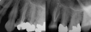
Radiographs of tooth 10 showing persistent apical periodontitis five years after initial endodontic treatmentNewsletter March ’08 – The attached case illustrates how useful an long term application is of an intra canal medicament in treating persistent post treatment apical periodontitis in an endodontically well treated tooth. Patient received endodontic treatment by a very competent dentist 5 years ago in tooth 10. However, the tooth developed apical periodontitis subsequently.

Patient was referred to my office for apical surgery. My approach in these cases is to retreat the tooth whenever possible. In this case, the bridge was temporarily cemented, so gaining orthograde access to root canal system was not an issue.

IRecall radiographs at one, two and four months showing bony healing during long-term calcium hydroxide therapy retreated the tooth with calcium hydroxide as intracanal medicament for 5 months. As you can see from the 1,2 and 4 month recall, there seemed to be bony healing and i felt confident of obturating at the fifth month. MTA (mineral trioxide aggregate) was used as an apical plug and Gp+sealer for the rest of the root. Tooth was restored and bridge was permanently cemented.

Long term CaoH in this case was my choice because:

  1. Final radiographs after MTA apical plug, obturation and permanent cementation of the bridgeVery well done initial treatment (atleast radiographically) which suggests the recalcitrant nature of bacteria in the root canal system.
  2. To get a sense of the outcome before final restorative treatment.
  3. Avoid periapical surgery.

Excellent healing is observed with this approach. patient is ecstatic that surgery could be avoided.

The key to endodontic success is thorough debridement and disinfection of the canal space, effective obturation of the canal systems and an immediate definitive coronal restoration that prevents leakage and loss of tooth structure. Studies have proven over and over that delaying the placement of a definitve coronal restorartion on an endodntically treated tooth results in higher failure.

In my practice, a majority of the endodontically treated teeth receive a definitve coronal restoration immediately after the completion of the obturation of the canals.Some of these restorations involve a post placement in one of the bigger canals.

Post placement in my offcie is limited to:

Molars: if more than 2 walls are missing and if remaining dentin is less than what is required to retain the crown.

Premolars: more than one wall is missing, particularly if one of the walls is buccal or lingual and if remaining dentin is less than what is required to retain the crown.

Anteriors: if remaining dentin is less than what is required to retain the crown.

However, keep in mind that the single most important factor that influences positively the retention and the fracture resistance of a crown and the tooth respectively is the remaining dentin after the crown preparation. 2 mm solid dentin circumferentially (ferrule) is a critical necessity. Placing a post will not circumvent this necessity but only will compliment.

As you can see from the attached finished cases in the month of January, all teeth received definiteve restorations whether it is just sealing the access or doing a build up or post and core buildups. If consistency in performing high quality endodontic treatment defines a specialst endodontic practice, placing an immediate coronal buildup is a signature of excellence. Please review the last 40 cases that I finished in my practice defining this fact. Pre ops and at least one post op image is included.

History

  • 10 yr old girl.
  • Tooth 8 was fractured 3 weeks ago in a sporting event.
  • Patient was seen by her GP a week after the injury, who restored 8.
  • Case was referred to me for consult to evaluate 8 for endo.

Treatment

The tooth tested WNL to pulpal and periradicualar testing. so, no endodontic treatment was recommended. I noticed the pustules on her lip and careful history taking revealed the pustules appeared two weeks after the traumatic incident. The broken fragment was also not found after the trauma. I suspected the fragment could have been lodged in the lip and took a radiograph of the lip which showed the fragment. (radiograph attached). Surgery was performed to remove the fragment.

Relevant points

In any trauma case Its important to:

  1. Examine the facial structure for any splinters or foreign body or tooth fragment lodged
  2. Look for any fractures elsewhere in the facial skeleton.
  3. Perform vitality tests on all anterior teeth. You will be surprised to see how many pulps turn non vital in the long run even though there was no sign an injury at the moment of the traumatic incident.
  4. Reviews to assess vitality.
  5. Update yur knowledge with the latest guidelines of trauma management. have a copy ready in the treatment rooms.

Cases like these are not very common, but when they happen, its important to diagnose correctly and treat quickly.

Quite a few colleagues ask me if I do all my treatments in one visit and if I do a case in multiple visits, what are the criteria for multiple appointments? While it’s true that a lot of cases that I do are done in a single visit (which includes a permanent core build up) I still do quite a few cases in 2 or more visits. The reasons for 2-visit endo are:

Cases with necrotic pulp and large lesions, I tend to do them in 2 visits (even if the canals are dry at the end of visit 1)

  1. Canals that cannot be dried at the end of first visit.
  2. Retreatment of cases with lesions.
  3. Vital cases with a lot of pulp tissue in the isthmus that bleed incessantly.
  4. Emergency cases that have to be worked into the busy schedule with not enough time for a complete endodontic treatment.
  5. When the case is anatomically challenging and needs more time than what one appointment can afford us.
  6. Patients that cannot open their mouth for full appt time.
  7. Questionable perio/endo cases that need a two visit approach to see if the perio symptoms disappear after the first visit.
  8. Questionable endo cases (read : cases with treatment, retreatment, surgery done previously) that I dont want to commit the patient to a full fee until I know my treatment is getting the healing response.
  9. Trauma cases where teeth were avulsed/ luxated extruded intruded and teeth with open apices.

These are the general guidelines that I follow. once a while there may be exceptions to the rule and I may decide to do some of the above mentioned cases in one visit.

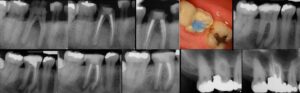
Here are the last few cases that I treated in my practice past 2 to 3 weeks. Some of them are done in 2 visits and some are in one.

You see the pattern that I followed with these cases. You can also appreciate the complex anatomy of the cases that we have here and the need to address this anatomy in a predictable and consistent basis. My mentor Dr.Gary Carr says “you are only as good as your last root canal”. I like to say, “‘you are only as good as your last 10 root canals”. Consistency is the key to endodontic excellence.

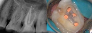
Here are the 4 maxillary molars that I treated in my practice today. 3 are second molars and 1 first molar. All teeth had 4 canals. 3 of the teeth had mb1 join mb2. One had 2 separate canals. All the cases were done in 1 visit. All teeth had self cure resin cores placed in the same visit to prevent coronal leakage and prevent tooth loss.

Many patients , once they are out of discomfort , will delay the placement of crowns leaving temporary fillings in endo treated teeth for more than a 2-4 weeks is not a good practice. My protocol is once the patient is anesthetised, apply rubber dam, remove decay, and any undermined tooth structure. Make my access into the chamber (if one exists) identify all canals, extend my coronal access into the canals to create straight line access to the canals. Use full strength bleach and 17% EDTA as irrigants. I use typically 20 ml of bleach for each canal and 5 ml of EDTA for each canal.

I typically use the irrigants between each file. I use NITI rotary instruments for bulk shaping of the canal and a lot of hand .02T hand instruments to fine tune the apical perparations. I use electronic Apex locator to determine the length of the canals. Of course, sometimes I take graphs/cone fit images to verify length and more so to rule out additional anatomy in the canals. So I like to call them working wire images than working length images.

Once the canals are fully shaped, I fit tapered cones usually .08T tapered cones. Once the canals are dried, I use paperponts to verify my length and then trim the cones to the final lengths. Warm vertical obturation is the choice of my obturation. I use Touch N Heat to do a down pack to at least 4-5 mm from the apex and then back fill with Obtura gun. Then clean off the chamber with alcohol, EDTA and then etch, bond and retore with self cure or dual cure resin and very often amalgam all under the rubber dam.

The whole procedure , once the rubber dam goes on, to it comes off, is done under a microscope. This allows the treatment to be done very precisely, find all the canals through the chamber calcifications, deep exploration of the canal spaces for deep splits, removing separated instruments, bypassing the ledges. The overall quality of the treatment improves significantly if the scope is used for the whole procedure. Quaity endodontics takes time, commitment and an attitide for excellence.

The results will speak for themselves when these criteria are met. No one does a perfect case every time, but consistency can be achieved if these principles are followed.

Tooth 20 was referred for endodontic retreatment. Patient has increased discomfort chewing and also spontaneous pain. Dx: Acute apical periodontitis with atleast 2 root canals untreated.

End of visit 1, all canals were cleaned and shaped to length. 2 buccal canals (MB,ML) and 1 Lingual canal were identified and treated. MB canal was the hardest of all to treat as the original treatment has ledged the canal space making negotiating to length difficult.

2nd visit – The canals were obturated and I placed cavit /IRM as interim restoration. patient is returning for retreat in 19 and the final restoration wll be done at that time.

Relevant Facts: Up to 40% of lower bicuspids have more than 1 canal. Even more so in black populations. Only 5% of all lower bicuspids have 3 canals and that is the highest number reported in the literature. These cases are fun to treat if one uses magnification and special microendodontic instruments. Otherwise its very stressful to treat these teeth. When 3 canals exist more often than not there are 2 canals to the buccal and 1 to the lingual.

Occasionally they could lie in a Letter \’C\” shape. its important to recognise the anatomy from straight and angled radiographs. case selection is key in these cases if you want to avoid misadventures.

Hope you find this case interesting.