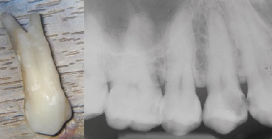
Most maxillary premolars have one or two root canals (Pathways of the Pulp, 8th edition), but a small percentage have three. One study reported three canals in 6% of the cases studied (Cairns and Skidmore, 1997).
If one is to treat three-canal maxillary premolar teeth predictably, it is necessary to be aware of the clinical and radiographic anatomy. These teeth may also require special shaping and obturating techniques.
A genetic component exists in the configuration of tooth forms. Three-canal maxillary premolars are fairly common in certain patient populations. In the author’s private practice in Jamaica, which consists largely of black Jamaicans and Jamaicans of mixed ethnicity, treatment of these teeth is a frequent occurrence. Over 25% of the maxillary first bicuspids the author has treated since the introduction of a surgical operating microscope to his practice have three canals.
The purpose of this article is to discuss how these teeth may be identified and the treatment modifications that are necessary for successful results.
Diagnosis: radiographic interpretation
Accurate preoperative radiographs using paralleling techniques are essential in providing clues as to the number of roots that exist (Silha RE, 1968).
In three-canal maxillary premolars, the cervical half of the root is generally wider than usual, with little or no taper.
Root canals may not be evident radiographically or may look unusual. Careful interpretation of the periodontal ligament space may suggest the presence of an extra root.
Mesial and distal angled views will often reveal the presence of two buccal roots.
Diagnosis: clinical appearance
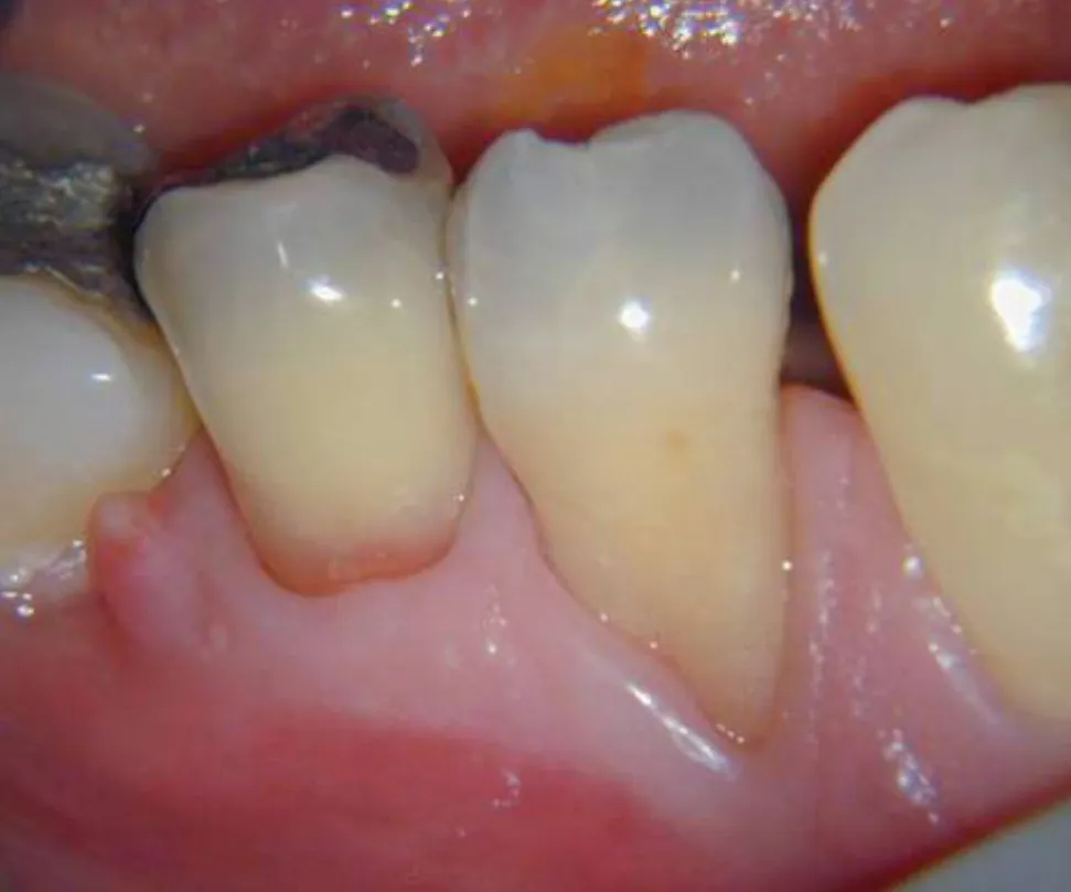
Clinical presentation is frequently atypical. A flattened buccal gingival collar is common, as opposed to a normal parabolic shape. This may reflect the furcal morphology in these teeth and, thus, hint at the presence of two buccal roots.
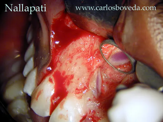
Probing the buccal sulcus to feel the root eminences and furcal anatomy may also help to identify the presence of two buccal roots. The use of magnification and fiber-optic illumination offers a tremendous advantage in locating and treating extra canals (Carr GB, 1983; Ruddle CJ, 1997). The dental operating microscope has been found to be particularly helpful by the author. Perhaps it is most important always to be on the look out for additional canals.
Tooth anatomy varies, but several features are commonly witnessed. In the author’s practice, there were two buccal canals and one palatal canal in every instance.
The division of the buccal canals was located either just above the cemento-enamel junction (most common) or in the mid-root area. For those teeth that separated in the mid root, it was necessary to enlarge the single buccal orifice and explore the mid-root.