The Significance and Use of Ophthalmic Dyes in the Location of Root Canal Orifices
Sashi Nallapati BDS and Gary Glassman DDS, FRCD(C) illustrate the significance and use of ophthalmic dyes in the location of root canal orifices
The relationship between uninstrumented root canals and endodontic treatment failure has been studied extensively. Locating all the canals, then shaping and cleaning them in their entirety has been shown to be essential for predictable clinical and biological success (Hoen M, Pink F, 2002; Siqueira JF Jr, 2001; Crump S, 1979; Cheung GS, 1996).
High visual magnification and fiber-optic illumination incorporated in the surgical operating microscope (SOM) has revolutionized endodontic therapy. The use of the SOM has facilitated the ease with which root canals are found in their typical, as well as aberrant, positions during orthograde endodontic treatment (Carr GB, 1998; Ruddle CJ, 1997).
Although the SOM is an indispensable aid in visualizing the detailed anatomy of the pulp chamber, it is essential to develop the visual acuity to appreciate the subtle differences that aid in the location of the root canals. The inherent color differences between the axial dentin and pulpal floor dentin, the coronal dentin and radicular dentin, as well as the differences in color and consistency between soft tissue and hard tissue, will assist in locating the root canal orifices (Niemczyk S, 1976).
This is of even greater significance in teeth with full coverage where the orientation markers of the natural tooth cannot be seen, such as cusp tips, grooves and the external contours of the root outlines. Other situations where the ‘pulpal road map’ has been altered are teeth that have been previously endodontically treated where canals have been missed, where prior occlusal access has been made, altering the chamber floor anatomy, and teeth with pulp chamber obliterations and canal calcifications.
Any help in terms of ‘marking’ the pulp tissue in canal orifices will facilitate the location of canals in both conventional and retreatment cases.
It is the purpose of this article to illustrate the significance and use of ophthalmic dyes in the location of root canal orifices (Niemczyk S).
Ophthalmic dyes
Ophthalmic dyes (e.g. fluorescein sodium, rose bengal) are currently being used in ophthalmological diagnostic procedures and for locating damaged areas of the cornea due to injury or disease.
Other uses for these dyes in ophthalmology include detection of epithelial defects, evaluation of the nasolacrimal system, determination of tear breakup time, angiography, location of non-epithelialized foreign bodies and contact lens pressure points (Newell FW, 1986).
Fluorescein sodium is available in pharmacies as a clear, orange-red solution as sterile, single-dose disposable eye drops in cartons of 10 units. Each unit contains approximately 0.5 ml. It is also available as individual strips. When the strips are used, the agents can be reconstituted by immersion in a dappen dish that contains sterile water or 90% alcohol (Figures 1 and 2). There are no serious contraindications reported for its use topically, except possible hypersensitivity. No serious side effects have been reported except for nausea (Newell FW, 1986).
There are few references for the use of ophthalmic dyes and fluorescence in dentistry. Those that are of significance have studied the use of ultra-violet induced fluorescence spectroscopy in diagnosis, pulp and root canal location, as well as using fluorescent spectroscopy to measure the relative sealing efficiency of root canal sealers (Foreman PC, 1983; Pini R et al, 1989; Taher M et al, 1973).
How they work
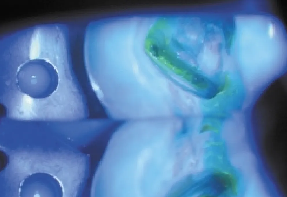
When these dyes come into contact with vital or non-vital pulp tissue they are readily absorbed by the connective tissue elements of the pulp in the chamber and root canal system. When exposed to blue light, these dyes dramatically fluoresce, showing scattered tissue segments that contrast with the surrounding monochromatic dentin. It is this quality that makes them useful in the location of pulp tissue in root canals.