
A maxillary right lateral incisor with a type 3 dens invaginatus and a large periapical lesion with vital pulp in a separate root canal was treated both nonsurgically and surgically. Care was taken not to expose or devitalize the vital pulp in the main root canal system during the treatment. The signs and symptoms ceased after the treatment, and 4-month recall showed complete bone healing with pulp vitality maintained.
Dens invaginatus is a rare malformation of teeth with a broad spectrum of morphological variations. The affected teeth present with an infolding of enamel and dentin, which may extend into the pulp cavity, into the root, and sometimes to the root apex (1). Several theories have been proposed for this phenomenon, but the etiology of dens invaginatus remains unclear. Kronfeld (2) proposed that dens invaginatus is caused by a focal failure of growth of the internal enamel epithelium leading to proliferation of the surrounding normal epithelium with eventual engulfment of the static area. Oehlers (3) proposed that distortion of the enamel organ occurs during tooth development and results in protrusion of a part of the enamel organ. Other theories include infection (4), trauma (5), and genetics (6) as possible contributing factors.
Oehlers (3) classified these malformations into three types. Of particular interest in his classification is type III, in which an enamel and/or cementum-lined dens forms tracks through the root and perforates in the apical area to form a second foramen but has no direct communication with the pulp. The purpose of this article is to discuss the clinical management of this particular variant of the dens invaginatus form.
CASE REPORTS
A healthy 24-year-old man was referred to the author’s private practice with the chief complaint of a gum boil above the upper right front tooth. The patient had no significant medical history.
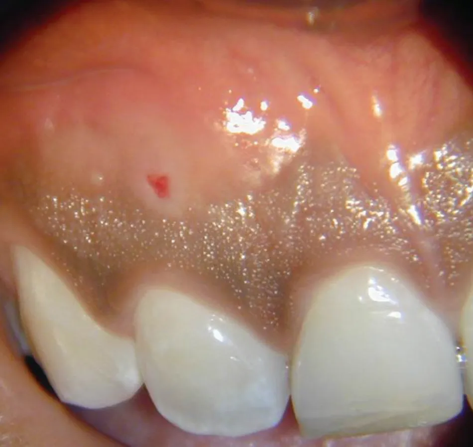
Clinical examination revealed an intraoral sinus tract in the labial gingiva adjacent to the maxillary right lateral incisor (Fig. 1). Clinical tests revealed the maxillary right lateral incisor to be within normal limits to percussion and slightly tender to palpation, with a normal response to cold. The vitality of the pulp was later confirmed with a test cavity. Radiographic examination revealed a $1 \times 2\text{ cm}$ radiolucency adjacent to the mesial aspect of the apex of the maxillary right lateral incisor (Fig. 2). The sinus tract was traced with a gutta-percha point to the lesion.
Radiographs revealed an enamel-lined tract, mesial to and separate from the main root canal system, that tracked to the apical radiolucency and appeared to be wide-open at the portal of exit. There was a separate root canal system distal to the dens that appeared to be closed apically. A diagnosis was made of normal pulp with chronic apical periodontitis associated with the type 3 dens invaginatus.
A treatment plan was formulated that included nonsurgical endodontic treatment of the dens tract over several appointments, including placement of a calcium hydroxide dressing. Surgical intervention was considered a strong possibility. Every effort would be made to leave the main root canal system undisturbed.
After a test cavity, which confirmed the vitality of pulp, the tooth was anesthetized, and nonsurgical endodontic treatment was initiated. All procedures were performed with the aid of a surgical operating microscope. Of particular interest was the atypical presentation of the lingual surface of the tooth. A rubber dam was placed, and access was made in the mesiolingual aspect of the tooth (Fig. 3). Upon access into the dens, there was a serosanguinous discharge, which was allowed to drain (Fig. 4). The canal was shaped initially with long tapered diamond bur to improve access to the apical defect. Working length of the dens tract was determined with the help of an apex locator (Root Zx, J Morita). The dens was debrided as well as possible with Gates Glidden drills and hand files. Because the canal was lined with enamel, methods for cleaning and shaping were not very effective. Irrigation was performed with 5.25% sodium hypochlorite, 17% EDTA, and 100% alcohol. Calcium hydroxide (Ultracal, Ultradent, UT) was placed in the dens tract between appointments (Fig. 5). The access cavity was sealed with Cavit and IRM between visits to prevent contamination of the dens canal system.


