Successful endodontic treatment involves accurate diagnosis, good understanding of the biological principles and excellent execution of the treatment. To be able to execute an excellent treatment, it’s imperative that the clinician has comprehensive knowledge of the root canal anatomy and the know-how to locate and treat this anatomy.
Many outcome studies conducted over the past few decades showed incomplete debridement and disinfection of root canal space as the most important factor in endodontic treatment failure (1,2,3). Missed canal in the initial treatment as a significant cause of root canal failure was shown by Hoen et al (4). They also showed a significant relation between an asymmetrical obturation in a root space and the incidence of a missed canal in an initial treatment.
Methodology
Root canal anatomy is studied by both in vitro and in vivo methods. In vivo methods include clinical treatment of a tooth followed by radiographic evaluation of the root canal anatomy. In vitro methods include:
- Direct observation
- Microscopic observation
- Macroscopic sectioning
- Microscopic sectioning
- Dyes
- Filling and decalcification
- Filling and clearing
- Radiography
- Contrasting media (Hypaque)
- Cone beam Tomography
Classification
Weine classified root canal anatomy into 3 types:
- Type I: One canal with one orifice and one apical foramen (1-1)
- Type II: Two canals that merge into one and exit as one canal (2-1)
- Type III: One canal that divides into two and exit as two canals. (2-2)
Vertucci’s classification was more elaborate and it covered 8 types:
- Type I: 1-1
- Type II: 2-1
- Type III: 1-2-1
- Type IV: 2-2
- Type V: 1-2
- Type VI: 2-1-2
- Type VII: 1-2-1-2
- Type VIII: 3-3
Studies with Percentages
The methodology employed and the criteria used to describe root canal anatomy will decide the percentages of canals found in any tooth. For example, Neaverth et al (5) have used “two separate canals could be visualized on radiographic examination (two files or two GP points to no less than a mm short of the length)” as the criteria for two canals in a mesiobuccal root of a maxillary first molar. On the other hand, Sempira et al (6) have used the presence of “two canals to at least 4 mm from the apex” in the mesiobuccal root of maxillary first molar to determine the percentage of Mb2 canal.
These differences in criteria dictate the great variation seen in the percentages of root canals seen in different studies. However, if one reviews the published evidence, certain features of aberrant anatomy stand out in a tooth/root.
- Ethnicity has a significant influence on aberrant anatomy (26). Radix Entomolaris, an extra distal root in a mandibular molar, is often seen in Oriental and Eskimo populations (23). Similarly, 2 and 3 canal premolars are seen frequently in black populations (12,15,27). ‘C’ shaped anatomy is seen more commonly in Chinese, Korean and Indian populations (22,25).
- Bilateral symmetry is a feature of aberrant anatomy. Rarer the aberration, the more common is the bilateral symmetry (28).
Clinical Management of Aberrant Anatomy
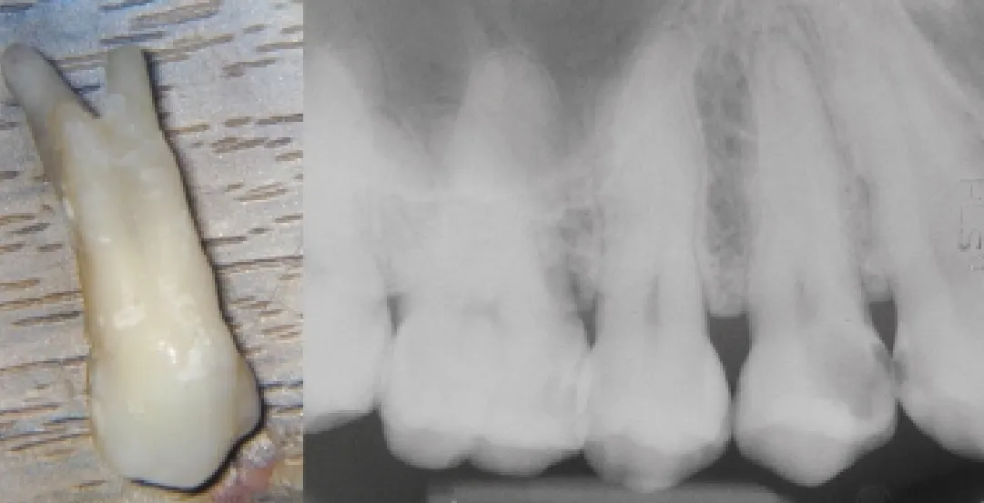
Radiography: Angled views of teeth reveal aberrant anatomy. Angled views allow us to visualize the root anatomy in 3 dimensions so that better assessment of the root canal anatomy is made. It is imperative that at least 2 angled views shall be taken before attempting endodontic treatment.
Author Profile
Dr. Sashi Nallapati obtained his dental degree from the Govt. Dental College and Hospital, Hyderabad, India. He completed his post graduate training in the specialty of Endodontics from Nova Southeastern University (NSU), Davie, Florida, USA. He maintains a practice limited to Endodontics in Kingston, Jamaica. He serves on the faculty of NSU in the dept. of Post Graduate Endodontics. Dr. Nallapati authored several clinical articles and lectures across the globe. His hobbies include digital photography, swimming and reading. He can be reached atwww.endojamaica.com.